The Beall Innovation Award in the Physical Sciences
The Beall Innovation Award in the Physical Sciences, launched in 2014, is the culmination of the vision and philanthropy of the Beall family. While funding agencies typically fund proposals aimed at advancing our understanding of science, the unique goal of the Beall Innovation Awards in the Physical Sciences is to promote the translation of science to commercialization and ultimately, to societal benefit.
Professor Jenny Yang, of the Department of Chemistry, has received the 2024 Award, marking the ten-year anniversary of the Beall Innovation Award in the Physical Sciences. Her proposal and presentation, titled "Exhale: CO2 Capture and Concentration from Indoor Air" describes recent technology from her research group that enables the ultra-efficient electrochemical removal of CO2 from indoor air. This innovation has the potential to dramatically reduce the expense associated with rapid air turnover in homes and businesses.
The decade-long support of this Award by the Beall family has been tremendously impactful for the School and the more than twenty faculty (listed below) who have received the Award since 2014. Updates from several recent award winners are also provided below.
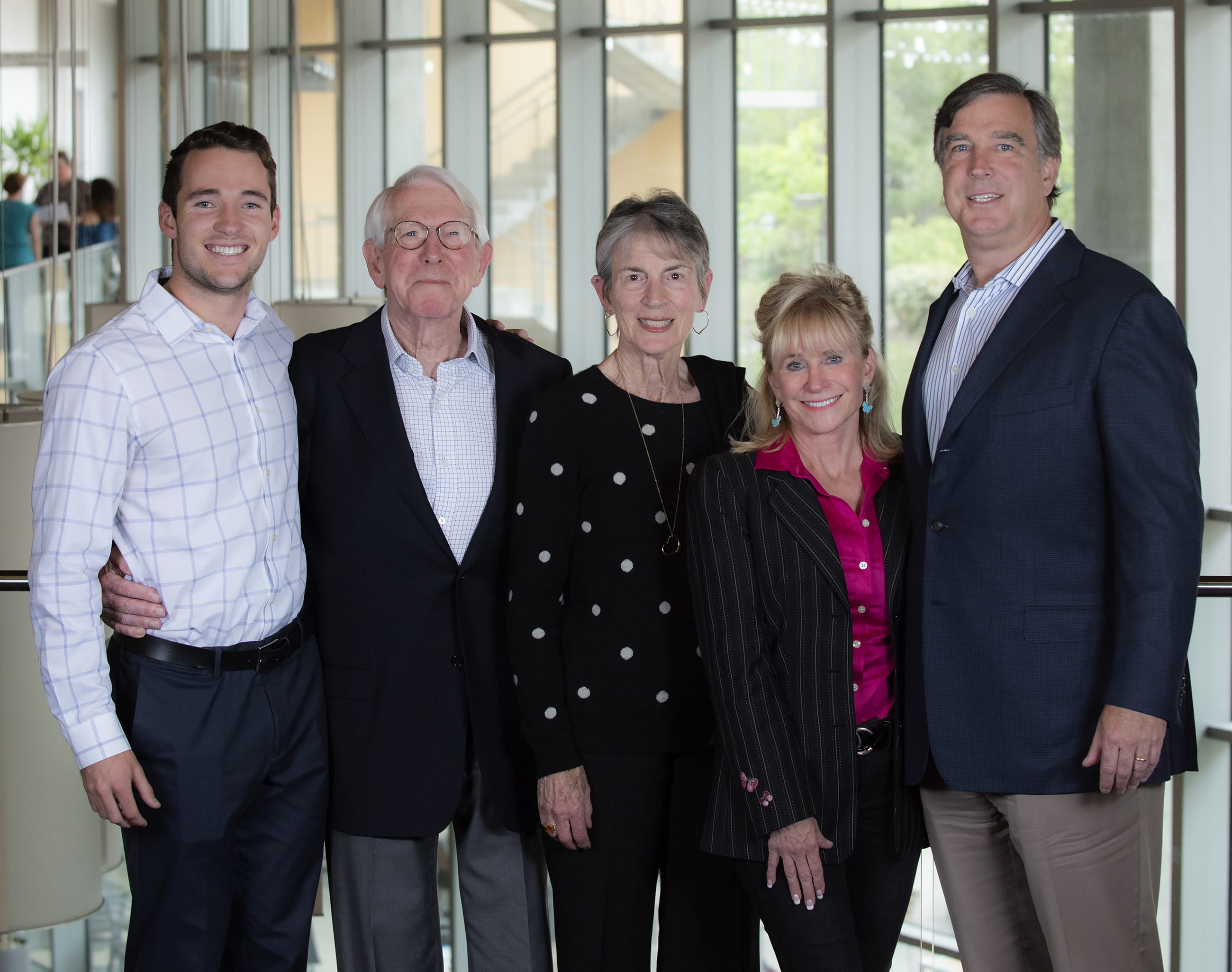
The Beall Family: (L to R) Charlie Beall, Don Beall, Joan Beall, Cher Beall, Ken Beall. Photo credit: Steve Zylius / UCI
Table of Recipients of the Beall Innovation Award in the Physical Sciences
Year | Scientist(s) | Project Title(s) |
|---|---|---|
2014 | Suzanne Blum | Commercialization of New Building Blocks for Drug Discovery. |
Zhibin Guan | Multiphase Self-Healing Polymers for Protective Coating Applications. | |
AJ Shaka | Nuclear Waste Recycling. Method to determine isotopic composition via delayed neutron spectroscopy with various foil surrounds. | |
2015 | Rachel Martin | NMR Probe Technology, proof-of-product development stage from proof-of-principle working 4-channel magic angle spinning probe |
Reg Penner & | Bioelectronic Devices. Use phage display to isolate receptors for recognition of new cancer biomarkers. | |
Ken Shea | Coating or Polish Removable by an Aqueous Solution of Cysteine. Development of self-immolative polyurethane polymers containing cysteine-activated trigger. | |
Zuzanna Siwy | Resistive-Pulse Sensing of Particles and Cells. Microfluidic device for electrical signal and high-speed camera to identify viable cancer cells under deformation. | |
2016 | Ken Shea | Broad spectrum abiotic antivenom against venomous serpent bites. |
Zuzanna Siwy | Nanopore 3D membranes. Membranes with pores which may discriminate cellular or ionic flow in microfluidic channels. Applications conceived in cancer stem cell isolation and also water desalination. | |
David Van Vranken | Antifungal drug compounds. | |
2017 | Peter Taborek | Trace quantities explosives detector |
Shane Ardo | Ion-Exchange Membrane: Fuel cells, Electrolyzers, | |
2018 | Peter Taborek(Physics & Astronomy) | Adaptive online learning platform for introductory physics |
Ilya Krivorotov & | Magnetic Field Effect Transistor for Drug Delivery | |
2019 | Eric Potma | Novel microscope objective lens for all colors. |
2020 | Shane Ardo | Ratchet-based ion pumping membrane systems. |
Joe Patterson | Artificial intelligence microscopy (AIM) for | |
2021 | Huolin Xin | Toward commercialization of compositionally complex doped ultra-high-nickel layered cathode for |
2022 | Jennifer Prescher | Smartphone detection of bioluminescent phasors |
Albert Siryaporn | Use of mammalian histones to establish | |
2023 | Howard Lee | Electrically tunable “meta”-optical fiber endoscopes for revolutionizing colonoscopy |
Highlights from Recent Award Recipients
Jennifer Prescher (Chemistry) – 2022 – Title: Smartphone detection of bioluminescent phasors for point-of-care diagnosis.
The Prescher group was awarded a 2022 Beall Award for the development of a multiplexed point-of-care biosensor for the detection of diseases using bioluminescent “phasors” that recognize and bind multiple disease markers in blood.
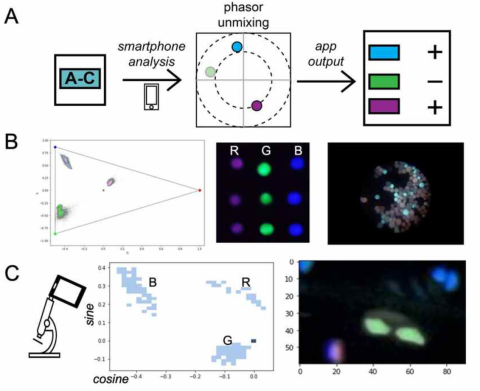
The scheme is shown schematically in Figure 1. They have been able to develop an easily accessible, smartphone detection platform for measuring bioluminescent phasors (Figure 1A). Custom bioluminescent probes were crafted and used to generate a modular workflow. As shown in Figure 1B, the method was readily able to distinguish mixtures of three different probes. They were further able to integrate the smartphone platform with easily deployable microscopes, to detect bioluminescent phasors in single cells. This work sets the stage for multiplexed analyses of bioluminescent sensors relevant to a variety of physiological processes. They are currently examining protease-specific sensors that can provide readouts on infectious disease.
The Prescher group is also preparing a publication on the design and construction of the smartphone platform including initial benchmarking results. They anticipate submitting the work this summer, along with a provisional patent application. In progress are the development of custom reagents and smartphone workflow for undergraduate education activities within the School of Physical Sciences and Engineering.
Albert Siryaporn (Physics and Astronomy) – 2022 - Title: Use of mammalian histones to establish a new class of antimicrobial treatments.
Professor Siryaporn received a Beall Award in 2022. This award has enabled them to assess the efficacy of our synergistic antimicrobial approach, which combines histone H2A and the antimicrobial peptide (AMP) polymyxin B, in a living organism (in vivo). They have assayed the activity of treatment in a corneal infection assay. Previous experiments in their lab showed that the combination of these molecules produced strong antimicrobial synergy due to the activity of AMPs enabling uptake of histones into the bacterial cytoplasm, where histones interfere with bacterial transcription and replication (Fig. 1A).

Their new work enabled by the Award shows that this treatment is effective in vivo against the opportunistic pathogen Pseudomonas aeruginosa and is a promising avenue of treatment of eye infections (Fig. 1B). The data shown in Figure 1 below is being incorporated into a manuscript that is in preparation. These data demonstrate that one of the challenges of this treatment is that the histones were removed rapidly after delivery due to the mouse eyelid motion. We have devised a new method to improve retention of the treatment using chitosan-embedded hydrogels. Their next step will be to increase retention of the histone on the corneal surface, and if successful, this will enable pre-clinical trials can be pursued to demonstrate greater efficacy (Fig. 1C). The Beall Innovation Award in the Physical Sciences has greatly assisted with his efforts to translate this research to commercialization.
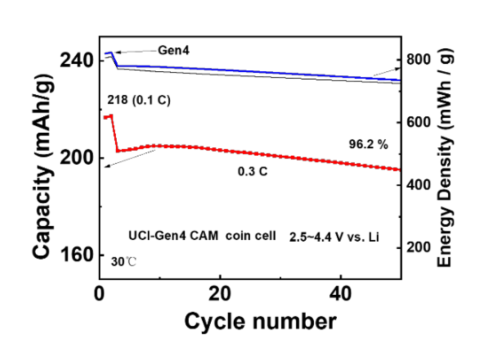
Huolin Xin (Physics) – 2021 – Title: Toward commercialization of compositionally complex doped ultra-high-nickel layered cathode for advanced lithium-ion batteries.
Professor Xin was awarded a Beall Award in 2021 for his proposal involving the development of a lithium ion battery cathode that contains zero cobalt. In this “Gen4 cathode”, nickel replaces all of the cobalt.
The development of a cobalt-free cathode represents a major break-through for lithium ion battery technology and we are proud that this has occurred in the Department of Physics and Astronomy at UCI!
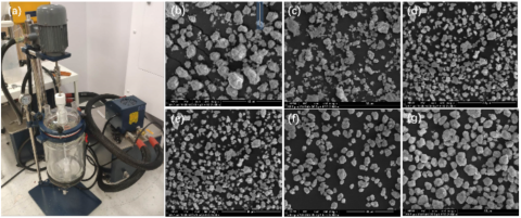
Now the challenge is to scale the synthesis of the Gen4 cathode from 50 g per batch to 200 g per batch via the installation of a 5-liter batch reactor (Fig. 1(a)). In addition, morphology control of the precursor was achieved through a series of experiments. Both rotation speed and concentration gradients were investigated to explore their effects on morphology. The results indicated that a higher rotation speed leads to a smaller particle size and a more homogeneous size distribution (Fig. 1(b-d)).
Using these optimized materials, Gen4 cathodes prepared at UCI have demonstrated a higher energy density compared to earlier products. The results (Fig. 2) show that the capacity retention by the battery is greater than 96% after 50 cycles – performance approaching conventional cobalt -containing cathodes now in use. The Xin group is seeking further improvements in cathode performance in the next year, based upon further optimization of the cathode synthesis process. Discussions with battery companies relating to this technology are in progress.